Hemodynamic property incorporated brain tumor segmentation by deep learning and density-based analysis of dynamic susceptibility contrast-enhanced magnetic resonance imaging (MRI)

Abstract
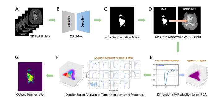
Background: Magnetic resonance imaging (MRI) is a primary non-invasive imaging modality for tumor segmentation, leveraging its exceptional soft tissue contrast and high resolution. Current segmentation methods typically focus on structural MRI, such as T1-weighted post-contrast-enhanced or fluid-attenuated inversion recovery (FLAIR) sequences. However, these methods overlook the blood perfusion and hemodynamic properties of tumors, readily derived from dynamic susceptibility contrast (DSC) enhanced MRI. This study introduces a novel hybrid method combining density-based analysis of hemodynamic properties in time-dependent perfusion imaging with deep learning spatial segmentation techniques to enhance tumor segmentation. Methods: First, a U-Net convolutional neural network (CNN) is employed on structural images to delineate a region of interest (ROI). Subsequently, Hierarchical Density-Based Scans (HDBScan) are employed within the ROI to augment segmentation by exploring intratumoral hemodynamic heterogeneity through the investigation of tumor time course profiles unveiled in DSC MRI. Results: The approach was tested and evaluated using a cohort of 513 patients from the open-source University of Pennsylvania glioblastoma database (UPENN-GBM) dataset, achieving a 74.83% Intersection over Union (IoU) score when compared to structural-only segmentation. The algorithm also exhibited increased precision and localized predictions of heightened segmentation boundary complexity, resulting in a 146.92% increase in contour complexity (ICC) compared to the reference standard provided by the UPENN-GBM dataset. Importantly, segmenting tumors with the developed new approach uncovered a negative correlation of the tumor volume with the scores in the Karnofsky Performance Scale (KPS) clinically used for assessing the functional status of patients (−0.309), which is not observed with the prevailing segmentation standard. Conclusions: This work demonstrated that including hemodynamic properties of tissues from DSC MRI can improve existing structural or morphological feature-based tumor segmentation techniques with additional information on tumor biology and physiology. This approach can also be applied to other clinical indications that use perfusion MRI for diagnosis or treatment monitoring. Keywords: Brain tumor; segmentation; dynamic susceptibility contrast (DSC); perfusion magnetic resonance imaging (perfusion MRI); deep learning